| Home | E-Submission | Sitemap | Editorial Office |  
J Minim Invasive Spine Surg Tech > Volume 8(2); 2023 > Article
Lee, Shin, Yi, Ha, Kim, and Lee: Overview and Prevention of Complications During Biportal Endoscopic Lumbar Spine Surgery

Abstract

Unilateral biportal endoscopic spine surgery (UBESS) is a minimally invasive surgical technique that has gained popularity for its potential benefits in various spinal lesions. It involves 2 small incisions, providing wide and clear endoscopic visualization, and causes less soft tissue damage than open surgery. UBESS offers flexibility and versatility in approaching different spinal disorders, including decompression of the spinal cord and root in the cervical or thoracic spine, as well as lumbar discectomy and spinal stenosis. One of the strengths of UBESS is its similarity to microscopic techniques, allowing for 2-handed endoscopic surgery. This familiarity makes it easier for surgeons to adopt endoscopic techniques and overcome the learning curve associated with spine endoscopy. However, some potential complications are associated with biportal endoscopic spine surgery, including dural tear, epidural hematoma, infection, incomplete surgery, and neural injury. Although the overall incidence of these complications is relatively low, it is important for clinicians to be aware of them and understand preventive methods.

INTRODUCTION

Minimally invasive treatments in spine surgery have significantly advanced in recent years. These procedures aim to reduce iatrogenic complications, postoperative discomfort, infection rates, and intraoperative blood loss. By preserving the posterior motion segments and paraspinal muscles, they minimize hospital stays, promote faster healing, and enable quicker return to normal daily activities. Unilateral biportal endoscopic spine surgery (UBESS) has emerged as a minimally invasive technique that has shown clinical effectiveness and safety. It has gained popularity for its potential benefits in various spinal lesions. UBESS involves 2 small incisions, providing wide and clear endoscopic visualization and causing less soft tissue damage. As an emerging endoscopic technique, UBESS offers flexibility and versatility in approaching many spinal disorders, including decompression of the spinal cord and root in the cervical or thoracic spine, as well as lumbar discectomy and spinal stenosis. Another advantage of UBESS is the ability to perform 2-handed endoscopic surgery, similar to microscopic techniques. This familiarity facilitates the adoption of endoscopic techniques and helps surgeons overcome the learning curve associated with spine endoscopy. However, there are potential complications associated with biportal endoscopic spine surgery (Table 1). A meta-analysis by Liang et al. [1] reported an overall complication rate of 5%, with dural tear being the most common complication at 2%, followed by epidural hematoma with an incidence of 1%. While the overall incidence of these complications is relatively low, it is important for clinicians to be aware of them and understand preventive methods.

COMPLICATIONS OF UBESS AND THEIR PREVENTION

1. Dura Tear

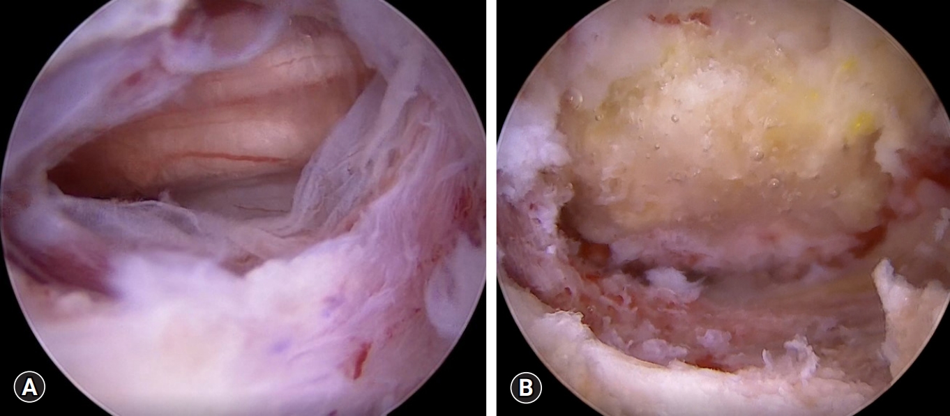
Dural tears are the most common complication in UBESS and have an incidence rate of 1.6%–14%. According to Liang et al. [1], dural injury was reported as the most common complication of UBESS for spinal stenosis, with a prevalence of 2%. During the unilateral laminotomy bilateral decompression procedure, the most common site of dural tear is the dorsal aspect of the dural sac, occurring during the removal of the ligamentum flavum [2,3]. The meningovertebral ligament, a web-like anatomical structure connecting the dura to the lamina and ligamentum flavum on the dorsal side, plays a significant role in these tears [4,5]. This ligament is predominantly located in the midline and can take the form of thin strips or thick sheets [5] Insufficient dissection of this ligament from the dura can lead to dural tears. In UBESS, while hydrostatic pressure can help separate the dural sac from the ligamentum flavum, folding can occur at the midline due to the presence of the meningovertebral ligament, potentially damaging the dural sac [1]. Lee et al. [2] suggested the use of angled curettes to remove small strips between the ligamentum flavum and dura (Figures 1, 2)
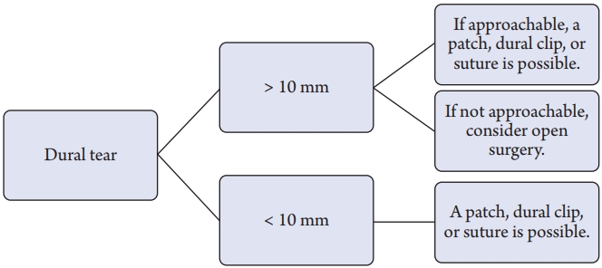
Dural tears may be associated with pseudomeningocele due to cerebrospinal fluid (CSF) leakage, surgical site infection, or rarely, meningitis. If dural repair is unsuccessful or not adequately treated, these complications can develop [6]. While open surgery typically involves primary repair as the standard treatment for dural tears, endoscopic spine surgery like UBESS does not have a standardized approach for dural tears. Kim et al. [7] proposed that small tears (<1 cm) can be effectively treated with the patch compression method, while large defects (≥1 cm) should be repaired using the dura clipping method. Choi et al. [8] suggested that minor tears (<4 mm) could be managed with bed rest alone, whereas larger tears (>12 mm) may require primary repair using a microscope (Figure 3).

2. Epidural Hematoma

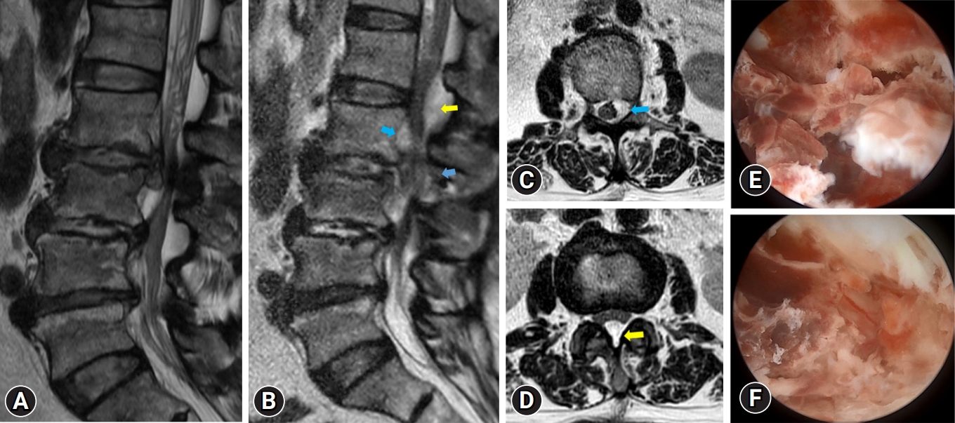
Postoperative epidural hematoma is a significant complication of UBESS as it is associated with postoperative infection, epidural fibrosis, or neurological compression [9,10]. In some cases, epidural hematoma can cause problematic compression of the spinal cord or cauda equina, resulting in a significant decline in patients' quality of life. Early recognition of symptoms is crucial for determining whether further evaluation and management are necessary. Symptoms of epidural hematoma include paralysis or bladder dysfunction at the spinal cord level, as well as intractable back pain or radicular pain at the lumbar level, usually occurring within 24 hours after surgery [11]. Mild postoperative hematoma symptoms without neurological deterioration typically resolve within 3 weeks after surgery, and radiologic regression occurs spontaneously within 3 months after surgery [12]. Several factors contribute to the development of epidural hematoma, including blood pressure control, postoperative drainage, preoperative anticoagulant or antiplatelet medication, and the use of intraoperative saline infusion pumps [13]. Fujiwara et al. [14] reported that patients with hypertension and poor blood pressure control experienced a more pronounced increase in blood pressure during extubation, which could lead to bleeding. Kim et al. [15] found that high water pressure ensures clear endoscopic visualization but may conceal bleeding from epidural vessels or bone.
Electrocoagulation is a common method used to control intraoperative bleeding. However, in cases where bleeding control is unsatisfactory, hemostatic materials such as microfibrillar collagen, thrombin gelatin, and gelatin-thrombin matrix sealant can be employed. Moreover, the use of bone wax for exposed cancellous bone or the insertion of a hemovac is a useful surgical tip to prevent epidural hematoma (Figure 4).

3. Incomplete Decompression

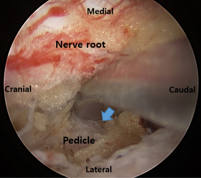
While decompression surgery with UBESS for spinal stenosis is usually excellent, in the case of severe lumbar spinal stenosis, decompression could be incomplete. Choi et al. [16] reported that inadequate resection of ligamentum flavum at the crainal and contralateral sides was related to patients experiencing radicular symptoms in their early cases. Choi et al. [16] suggested that angled curettes were more useful than Kerrison punches for performing a proper flavectomy. Angled curettes can scrape the ligamentum flavum under the lamina without requiring extensive laminectomy. To decompress the contralateral side, they recommended partial resection of the upper and lower ends of the spinous processes to create enough space for the insertion of the endoscope and instruments [16]. Moreover, the medial margin of the lower pedicle must be identified for ideal decompression of both nerve roots (Figure 5).
Blurred vision due to intraoperative bleeding can also contribute to incomplete decompression. Meticulous control of systolic blood pressure (below 100 mmHg) and the intermittent use of bone wax and gelfoam can help prevent this complication.

4. Recurrence

Recurrence after full endoscopic lumbar discectomy is associated with older age (over 50 yerars), obesity (body mass index > 25 kg/m2), higher lumbar disc herniation, and central disc herniation. Within 6 months, the disease history, Pfirrmann grade, Modic alterations, and migration grade can predict the total recurrence rate following endoscopic lumbar discectomy [17]. The aforementioned risk factors appear to be linked to recurrence of disc herniation. Soliman [18] described a case of recurrent disc herniation in a patient who had undergone UBESS.

5. Instability

Previous biomechanical investigations have found that laminectomies involving the excision of more than 50% of the pars interarticularis increase the likelihood of iatrogenic instability. Iatrogenic instability associated with UBESS could be linked to prolonged drilling of the facet joint, and excessive laminectomies are risk factors for this disorder. In a study by Kim et al. [15], the risk of iatrogenic instability was reported to be 0.6% because UBESS reduces muscle dissection and preserves the zygapophyseal joint compared to standard open surgery. In contrast, the rate of iatrogenic spondylolisthesis after open laminectomy is reported to be between 3.96% and 9.5% [19]. Iatrogenic instability can be avoided by undercutting the facet joint. It is critical to reduce facet joint infringement during surgery to prevent postoperative instability [20,21].

6. Root Injury

Radiofrequency (RF) probes are essential and widely used in UBESS. However, intraoperative thermal injury from RF has been identified as the primary cause of nerve root injury [1]. While direct contact thermal injury of the nerve root by the RF probe tip can be avoided through the surgeon's skill, indirect RF thermal injury resulting from the elevation of epidural temperature may not be entirely controlled by the surgeon [22]. Heo et al. [22] reported that RF can be safely used in UBESS, and the utilization of low-power and short-duration RF can reduce the possibility of thermal injury. Moreover, maintaining good irrigation patency in the surgical field is important for minimizing the elevation of epidural temperature caused by RF.
In UBESS, the use of a drill above the ligamentum flavum is safer than the use of a Kerrison punch to prevent root injury because ligamentum flavum can act as a barrier to protect the nerve roots during bone work.
When performing decompression at the L1/2 level, there is a possibility of spinal cord injury, particularly. Therefore, we must exercise caution to avoid compressing the thecal sac using surgical instruments such as retractors and Kerrison punches in the high lumbar segment area.

7. Infection

One notable aspect of UBESS is the absence of postoperative infection, which is a relatively common occurrence in conventional lumbar spinal surgery [23]. The incidence of spine infection after spine surgery ranges from approximately 0.1% to 4.5%, with bacterial infection being the most common cause [23]. However, UBESS has a low incidence of postoperative infection due to factors such as continuous saline irrigation, shorter operation time, and reduced soft tissue injury [24].

8. Postoperative Headache

In UBESS, the use of high intraoperative water pressure can increase CSF pressure and intracranial pressure, leading to postoperative headaches and, in severe cases, seizures [25]. Therefore, it is important to monitor patients for symptoms such as neck pain, headaches, blurred vision, and drowsiness. To prevent the occurrence of postoperative headaches, it is crucial to control intraoperative water pressure, fluid outflow, and operation time. Choi [26] advised keeping the irrigation pump pressure below 30 mmHg. Kang et al. [27] reported that cervical epidural pressure remains within the physiological range when continuous lavage is performed with an infusion pressure set to 30 mmHg. Kim et al. [28] suggested that extending the fascia incision of the working portal would be preferable to improve fluid outflow. Czigléczki et al. [29] reported that irrigation could cause meningeal irritation and postoperative headaches, but reducing the operation time can help avoid such complications.

9. Retinal Hemorrhage

After a UBE discectomy, Lee et al. [30] described a patient with retinal hemorrhage. They suggested that increased CSF pressure may have been responsible for the retinal bleeding during the unilateral biportal endoscopic (UBE) discectomy procedure. This pressure could have been transmitted to the retinal venous circulation either directly through the optic nerve sheaths or indirectly through the subarachnoid extension surrounding the optic nerve. Furthermore, higher CSF pressure has the potential to reduce cerebral blood flow, triggering a reflex increase in ophthalmic artery pressure, which can lead to capillary rupture and venous collapse. According to Lee et al. [30], it is crucial to regulate the pressure of the irrigated fluid during UBE to prevent rare complications such as postoperative retinal bleeding.

CONCLUSION

As a minimally invasive technique, UBESS has been successfully used for lumbar spine disorders and has gained popularity due to its therapeutic efficacy, including satisfactory clinical outcomes, shorter hospital stays and operation times, and lower complication rates. Based on a literature review, the most common complications of UBESS include dural tear, epidural hematoma, nerve root injury, incomplete decompression, and postoperative headache. It is crucial to have a comprehensive understanding of the procedure, surgical technique, complications, and prevention strategies associated with UBESS.

NOTES

Conflicts of Interest

The authors have nothing to disclose.

Funding/Support

This study received no specific grant from any funding agency in the public, commercial, or not-for-profit sectors.

Figure 1.
Dural tear. (A) Endoscopic view of dural tear (about 10 mm on the dorsal side). (B) Endoscopic view of dural repair with a fibrin collagen patch.
jmisst-2023-00885f1.jpg
Figure 2.
Dural fold and posterior epidural ligament. (A) Dural folding due to hydrostatic pressure (blue arrow). (B) Posterior epidural ligament or meningovertebral ligament (red arrow).
jmisst-2023-00885f1.jpg
Figure 3.
Treatment algorithm of dural tears.
jmisst-2023-00885f3.jpg
Figure 4.
A 65-year-old female patient with left leg pain and left leg weakness (G4-). After undergoing discectomy, her radicular pain disappeared and the leg weakness improved. However, 3 days after surgery, she experienced severe left leg pain and developed progressive leg weakness (G2). Magnetic resonance (MR) imaging revealed a postoperative hematoma at surgical site (L2/3), which extended to an upper level (L1/2). Following revision surgery, her radicular pain subsided and her leg weakness improved, but persisted. (A) Preoperative MR image shows herniated lumbar disc L2/3 left with spinal stenosis. (B) Postoperative MR sagittal image shows epidural hematoma (yellow and blue arrows). (C) Postoperative MR axial image shows epidural hematoma at L2 level (blue arrow). (D) Postoperative MR axial image shows epidural hematoma at L1/2 level (yellow arrow). (E, F) endoscopic view of epidural hematoma.
jmisst-2023-00885f4.jpg
Figure 5.
End point of lumbar foraminal decompression. The medial wall of the pedicle of the lower vertebrae is touched with a double ended dissector (blue arrow).
jmisst-2023-00885f5.jpg
Table 1.
Overview of complications of unilateral biportal endoscopic spine surgery in the reviewed study
Study Year Design Country Complications (case number)
Park et al. [3] 2020 RCT Korea Dural tear (2), hematoma (1)
Kim et al. [7] 2020 Retrospective Korea Hematoma (5), recurrence (16), dural tear (3)
Choi et al. [16] 2016 Retrospective Korea Dural tear (2), nerve root injury (1), incomplete decompression (1)
Eum et al. [25] 2016 Retrospective Korea Postoperative headache (3), dural tear (2), transient leg numbness (2), hematoma(1)
Czigléczki et al. [29] 2020 Retrospective Hungary Postoperative headache (3), dural tear (2), incomplete decompression (1)
Li et al. [31] 2022 Retrospective China Dural tear (1), transient paresthesia (1)
Jung and Kim [32] 2022 Retrospective Korea Transient motor weakness (1)
Zhu et al. [33] 2022 Technical note China Transient paresthesia (1)
An and Lee [34] 2019 Technical note Korea Operation site pain and numbness (1)
Lin et al. [35] 2019 Retrospective China Dural tear (1)
Kim and Park [36] 2020 Retrospective Korea Dural tear (2), root injury (3), infection (2)
Kim et al. [37] 2020 Retrospective Korea Dural tear (3), hematoma (1)
Kim et al. [38] 2019 Retrospective Korea Transient paresthesia (5)
Kang et al. [39] 2019 Retrospective Korea Dural tear (1)
Kang et al. [40] 2020 Retrospective Japan Dural tear (2)
Kim and Choi [41] 2018 Retrospective Korea Dural tear (2), hematoma (1)
Hong et al. [42] 2020 Retrospective Korea Dural tear (2)
Heo et al. [43] 2019 Retrospective Korea Dural tear (1), hematoma (1)
Heo et al. [44] 2018 Prospective Korea Dural tear (1), hematoma (1)
Pao et al. [45] 2020 Retrospective China (Taiwan) Dural tear (4), transient paresthesia (1), hematoma (1), incomplete decompression (1)
Song and Lee [46] 2020 Technical note Korea Dural tear (1)
Fishchenko et al. [47] 2020 Retrospective Ukraine Dural tear (4)
Ahn et al. [48] 2018 Retrospective Korea Dural tear (1)
Kim et al. [49] 2018 Retrospective Korea Incomplete decompression (3)
Eun et al. [50] 2017 Retrospective Korea Incomplete decompression (1)
Torudom et al. [51] 2016 Retrospective Thailand Transient paresthesia (2), incomplete decompression (1)
Soliman [52] 2015 Prospective Egypt Dura tear (6)
Min et al. [53] 2020 Retrospective Korea Dural tear (2), hematoma (1)

RCT, randomized controlled trial.

REFERENCES

1. Liang J, Lian L, Liang S, Zhao H, Shu G, Chao J, et al. Efficacy and complications of unilateral biportal endoscopic spinal surgery for lumbar spinal stenosis: a meta-analysis and systematic review. World Neurosurg 2022;159:e91–102.
pmid
2. Lee HG, Kang MS, Kim SY, Cho KC, Na YC, Cho JM, et al. Dural injury in unilateral biportal endoscopic spinal surgery. Global Spine J 2021;11:845–51.
crossref pmid pmc pdf
3. Park HJ, Kim SK, Lee SC, Kim W, Han S, Kang SS. Dural Tears in percutaneous biportal endoscopic spine surgery: anatomical location and management. World Neurosurg 2020;136:e578–85.
crossref pmid
4. Shi B, Zheng X, Min S, Zhou Z, Ding Z, Jin A. The morphology and clinical significance of the dorsal meningovertebra ligaments in the cervical epidural space. Spine J 2014;14:2733–9.
crossref pmid
5. Geers C, Lecouvet FE, Behets C, Malghem J, Cosnard G, Lengelé BG. Polygonal deformation of the dural sac in lumbar epidural lipomatosis: anatomic explanation by the presence of meningovertebral ligaments. AJNR Am J Neuroradiol 2003;24:1276–82.
pmid pmc
6. Kang SS, Kim JE, Choi DJ, Park EJ. Pseudomeningocele after biportal endoscopic spine surgery: a case report. J Orthop 2019;18:1–4.
crossref pmid pmc
7. Kim JE, Choi DJ, Park EJ. Risk factors and options of management for an incidental dural tear in biportal endoscopic spine surgery. Asian Spine J 2020;14:790–800.
crossref pmid pmc pdf
8. Choi DJ, Jung JT, Lee SJ, Kim YS, Jang HJ, Yoo B. Biportal endoscopic spinal surgery for recurrent lumbar disc herniations. Clin Orthop Surg 2016;8:325–9.
crossref pmid pmc pdf
9. Kanayama M, Oha F, Togawa D, Shigenobu K, Hashimoto T. Is closed-suction drainage necessary for single-level lumbar decompression?: review of 560 cases. Clin Orthop Relat Res 2010;468:2690–4.
pmid pmc
10. Kotil K. Closed drainage versus non-drainage for single-level lumbar disc surgery: relationship between epidural hematoma and fibrosis. Asian Spine J 2016;10:1072–8.
crossref pmid pmc pdf
11. Anno M, Yamazaki T, Hara N, Ito Y. The incidence, clinical features, and a comparison between early and delayed onset of postoperative spinal epidural hematoma. Spine (Phila Pa 1976) 2019;44:420–3.
crossref pmid
12. Ikuta K, Tono O, Tanaka T, Arima J, Nakano S, Sasaki K, et al. Evaluation of postoperative spinal epidural hematoma after microendoscopic posterior decompression for lumbar spinal stenosis: a clinical and magnetic resonance imaging study. J Neurosurg Spine 2006;5:404–9.
crossref pmid
13. Kim JE, Choi DJ, Kim MC, Park EJ. Risk factors of postoperative spinal epidural hematoma after biportal endoscopic spinal surgery. World Neurosurg 2019;129:e324–9.
crossref pmid
14. Fujiwara Y, Manabe H, Izumi B, Harada T, Nakanishi K, Tanaka N, et al. The impact of hypertension on the occurrence of postoperative spinal epidural hematoma following single level microscopic posterior lumbar decompression surgery in a single institute. Eur Spine J 2017;26:2606–15.
crossref pmid pdf
15. Kim W, Kim SK, Kang SS, Park HJ, Han S, Lee SC. Pooled analysis of unsuccessful percutaneous biportal endoscopic surgery outcomes from a multi-institutional retrospective cohort of 797 cases. Acta Neurochir (Wien) 2020;162:279–87.
crossref pmid pdf
16. Choi DJ, Choi CM, Jung JT, Lee SJ, Kim YS. Learning curve associated with complications in biportal endoscopic spinal surgery: challenges and strategies. Asian Spine J 2016;10:624–9.
crossref pmid pmc
17. Ogihara S, Yamazaki T, Inanami H, Oka H, Maruyama T, Miyoshi K, et al. Risk factors for surgical site infection after lumbar laminectomy and/or discectomy for degenerative diseases in adults: a prospective multicenter surveillance study with registry of 4027 cases. PLoS One 2018;13:e0205539.
crossref pmid pmc
18. Soliman HM. Irrigation endoscopic discectomy: a novel percutaneous approach for lumbar disc prolapse. Eur Spine J 2013;22:1037–44.
crossref pmid pmc pdf
19. Ahuja S, Moideen AN, Dudhniwala AG, Karatsis E, Papadakis L, Varitis E. Lumbar stability following graded unilateral and bilateral facetectomy: A finite element model study. Clin Biomech (Bristol, Avon) 2020;75:105011.
crossref pmid
20. Ramhmdani S, Xia Y, Xu R, Kosztowski T, Sciubba D, Witham T, et al. Iatrogenic spondylolisthesis following open lumbar laminectomy: case series and review of the literature. World Neurosurg 2018;113:e383–90.
crossref
21. Spina NT, Moreno GS, Brodke DS, Finley SM, Ellis BJ. Biomechanical effects of laminectomies in the human lumbar spine: a finite element study. Spine J 2021;21:150–9.
crossref
22. Heo DH, Park DY, Hong YH, Kim D, Kim JS. Temperature change of epidural space by radiofrequency use in biportal endoscopic lumbar surgery: safety evaluation of radiofrequency. Eur Spine J 2023;32:2769–75.
crossref pmid pdf
23. Kumagai G, Wada K, Asari T, Nitobe Y, Ishibashi Y. Association of methicillin-resistant coagulase-negative Staphylococci on preoperative skin and surgical site infection in patients undergoing spinal surgery: a retrospective cohort study. Spine Surg Relat Res 2022;6:596–603.
crossref pmid pmc
24. Ju CI, Lee SM. Complications and management of endoscopic spinal surgery. Neurospine 2023;20:56–77.
crossref pmid pmc pdf
25. Hwa Eum J, Hwa Heo D, Son SK, Park CK. Percutaneous biportal endoscopic decompression for lumbar spinal stenosis: a technical note and preliminary clinical results. J Neurosurg Spine 2016;24:602–7.
crossref pmid
26. Choi CM. Biportal endoscopic spine surgery (BESS): considering merits and pitfalls. J Spine Surg 2020;6:457–65.
crossref pmid pmc
27. Kang MS, Park HJ, Hwang JH, Kim JE, Choi DJ, Chung HJ. Safety evaluation of biportal endoscopic lumbar discectomy: assessment of cervical epidural pressure during surgery. Spine (Phila Pa 1976) 2020;45:E1349–56.
crossref pmid
28. Kim JE, Choi DJ, Park EJJ, Lee HJ, Hwang JH, Kim MC, et al. Biportal endoscopic spinal surgery for lumbar spinal stenosis. Asian Spine J 2019;13:334–42.
crossref pmid pmc pdf
29. Czigléczki G, Nagy Z, Padányi C, Banczerowski P. Biportal endoscopic technique in the treatment of spinal stenosis: early clinical experiences and results. Neurol Res 2020;42:1085–8.
crossref pmid
30. Lee KH, Kim GL, Park J, Lee HB, Hong SY, Kim TH. Retinal hemorrhage and transient consciousness disturbance after biportal endoscopic lumbar discectomy: a case report and literature review. J Orthop Sci 2021:2021 May 31:S0949-2658(21)00150-0. doi: 10.1016/j.jos.2021.04.013. [Epub].
crossref pmid
31. Li YS, Chen CM, Hsu CJ, Yao ZK. Complications of unilateral biportal endoscopic lumbar discectomy: a systematic review. World Neurosurg 2022;168:359–68.e2.
crossref pmid
32. Jung SB, Kim N. Biportal endoscopic spine surgery for cervical disk herniation: a technical notes and preliminary report. Medicine (Baltimore) 2022;101:e29751.
crossref pmid pmc
33. Zhu C, Cheng W, Wang D, Pan H, Zhang W. A helpful third portal for unilateral biportal endoscopic decompression in patients with cervical spondylotic myelopathy: a technical note. World Neurosurg 2022;161:75–81.
crossref pmid
34. An JW, Lee CW. Surgical treatment of extraforaminal gas-containing pseudocyst compressing L5 nerve root by using unilateral biportal endoscopy. World Neurosurg 2019;124:145–50.
crossref pmid
35. Lin GX, Huang P, Kotheeranurak V, Park CW, Heo DH, Park CK, et al. A systematic review of unilateral biportal endoscopic spinal surgery: preliminary clinical results and complications. World Neurosurg 2019;125:425–32.
crossref pmid
36. Kim KR, Park JY. The technical feasibility of unilateral biportal endoscopic decompression for the unpredicted complication following minimally invasive transforaminal lumbar interbody fusion: case report. Neurospine 2020;17(Suppl 1):S154–9.
crossref pmid pmc pdf
37. Kim HS, Wu PH, Jang IT. Lumbar endoscopic unilateral laminotomy for bilateral decompression outside-in approach: a proctorship guideline with 12 steps of effectiveness and safety. Neurospine 2020;17(Suppl 1):S99–109.
crossref pmid pmc pdf
38. Kim JE, Choi DJ, Park EJ. Evaluation of postoperative spinal epidural hematoma after biportal endoscopic spine surgery for single-level lumbar spinal stenosis: clinical and magnetic resonance imaging study. World Neurosurg 2019;126:e786–92.
crossref pmid
39. Kang T, Park SY, Lee SH, Park JH, Suh SW. Spinal epidural abscess successfully treated with biportal endoscopic spinal surgery. Medicine (Baltimore) 2019;98:e18231.
crossref pmid pmc
40. Kang MS, Hwang JH, Choi DJ, Chung HJ, Lee JH, Kim HN, et al. Clinical outcome of biportal endoscopic revisional lumbar discectomy for recurrent lumbar disc herniation. J Orthop Surg Res 2020;15:557.
crossref pmid pmc pdf
41. Kim JE, Choi DJ. Unilateral biportal endoscopic decompression by 30° endoscopy in lumbar spinal stenosis: technical note and preliminary report. J Orthop 2018;15:366–71.
crossref pmid pmc
42. Hong YH, Kim SK, Suh DW, Lee SC. Novel instruments for percutaneous biportal endoscopic spine surgery for full decompression and dural management: a comparative analysis. Brain Sci 2020;10:516.
crossref pmid pmc
43. Heo DH, Sharma S, Park CK. Endoscopic treatment of extraforaminal entrapment of L5 nerve root (Far Out Syndrome) by unilateral biportal endoscopic approach: technical report and preliminary clinical results. Neurospine 2019;16:130–7.
crossref pmid pmc pdf
44. Heo DH, Quillo-Olvera J, Park CK. Can percutaneous biportal endoscopic surgery achieve enough canal decompression for degenerative lumbar stenosis? Prospective case-control study. World Neurosurg 2018;120:e684–9.
crossref pmid
45. Pao JL, Lin SM, Chen WC, Chang CH. Unilateral biportal endoscopic decompression for degenerative lumbar canal stenosis. J Spine Surg 2020;6:438–46.
crossref pmid pmc
46. Song KS, Lee CW. The biportal endoscopic posterior cervical inclinatory foraminotomy for cervical radiculopathy: technical report and preliminary results. Neurospine 2020;17(Suppl 1):S145–53.
crossref pmid pmc pdf
47. Fishchenko I, Kravchuk L, Saponenko A, Roy I. Experience of biportal endoscopic decompression in lumbar spinal stenosis. Georgian Med News 2020;(303):21–7.

48. Ahn JS, Lee HJ, Choi DJ, Lee KY, Hwang SJ. Extraforaminal approach of biportal endoscopic spinal surgery: a new endoscopic technique for transforaminal decompression and discectomy. J Neurosurg Spine 2018;28:492–8.
crossref pmid
49. Kim SK, Kang SS, Hong YH, Park SW, Lee SC. Clinical comparison of unilateral biportal endoscopic technique versus open microdiscectomy for single-level lumbar discectomy: a multicenter, retrospective analysis. J Orthop Surg Res 2018;13:22.
crossref pmid pmc pdf
50. Eun DC, Lee YH, Park JO, Suk KS, Kim HS, Moon SH, et al. A comparative analysis of bi-portal endoscopic spine surgery and unilateral laminotomy for bilateral decompression in multilevel lumbar stenosis patients. J Clin Med 2023;12:1033.
crossref pmid pmc
51. Torudom Y, Dilokhuttakarn T. Two portal percutaneous endoscopic decompression for lumbar spinal stenosis: preliminary study. Asian Spine J 2016;10:335–42.
crossref pmid pmc
52. Soliman HM. Irrigation endoscopic decompressive laminotomy. A new endoscopic approach for spinal stenosis decompression. Spine J 2015;15:2282–9.
crossref pmid
53. Min WK, Kim JE, Choi DJ, Park EJ, Heo J. Clinical and radiological outcomes between biportal endoscopic decompression and microscopic decompression in lumbar spinal stenosis. J Orthop Sci 2020;25:371–8.
crossref pmid
Editorial Office
Department of Neurosurgery, Harrison Spinartus Hospital Chungdam
646 Samseong-ro, Gangnam-gu, Seoul 06084, Korea
TEL: +82-2-6003-9767    FAX: +82-2-3445-9755   E-mail: office@jmisst.org
About |  Browse Articles |  Editorial Policy |  For Contributors
Copyright © Korean Minimally Invasive Spine Surgery Society.                 Developed in M2PI