INTRODUCTION
Peroneal nerve palsy causes a weakness or paralysis of ankle dorsiflexion and eversion, as well as of the extensors of the toes. Peroneal nerve palsy is the most common cause of foot drop, with various mechanisms of injury having been identified, including direct trauma, tumor, infection and metabolic disorder [6,11,18]. Although less frequent, a foot drop can develop as a secondary outcome of a central lesion, such as anterior horn cell disease and/or lesion to the sciatic nerve, lumbar plexus, lumbo-sacral trunk and the axons of the L4 and L5 spinal nerves [15]. Although lower motor neuron and peripheral nerve lesions have commonly been described as causes of foot drop in clinical practice, a few studies have also reported lesions of the head and neck as possible causes of a foot drop, with stroke being a specific example of a central cause. The case study presented here describes the cause of a left foot drop that developed immediately after anterior cervical discectomy and fusion (ACDF) for herniation of the nucleus pulposus (HNP) at the spinal levels of C4-C5 and C5-C6.
CASE REPORT
A 67-year-old male patient presented to our outpatient clinic with complaint of left-side body numbness, which had been present for 1 year. Symptoms were aggravated with neck hyperextension. The distribution of the reported numbness was as follows: left upper limb, including the shoulder, forearm and all digits of the hand; trunk, extending from the umbilicus into the left buttocks; and the left calf. The patient also reported bilateral lower limb weakness, and could no longer run at cross-walks. He also reported unsteadiness, including a sensation of body sway during walking.
On neurological examination, the grading of muscle strength was 5/5 for both upper extremities; 4/5 for hip flexors, bilaterally; 4+/5 for hip adductors, bilaterally; and 4+/5 for all other muscle groups of the lower limbs, bilaterally. The patient was unable to stand on one foot, was unable to perform a single leg hop with either lower limb, and was unable to walk on heels or tip toe. Sensory examination identified paresthesia of the left palm and hyperesthesia of the lower limbs, bilaterally, including the feet. The Romberg and cerebellar function tests were positive, and Hoffman’s sign was positive for both hands, bilaterally. Hyperactive deep tendon reflexes were present at the knees and ankles, and the Babinski reflex was positive, bilaterally.
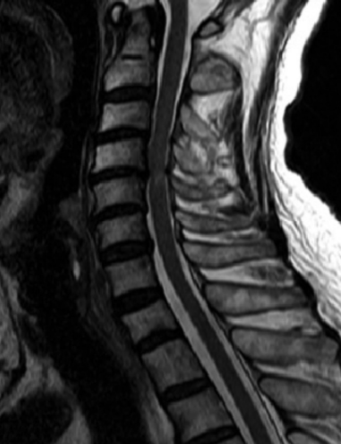
On magnetic resonance (MR) imaging of the cervical spine, diffuse cervical degenerative changes and black discs, indicative of complete dehydration and degeneration, were identified at all cervical levels. In addition, herniation of the nucleus pulposus (HNP), with thickening of the ligamentum flavum (yellow ligament), was also identified at C3-4 and C4-5 levels, predisposing factors of severe spinal canal stenosis. Change in MR signal of the spinal cord was also apparent at C3-C4 and C4-C5 levels (Fig. 1).
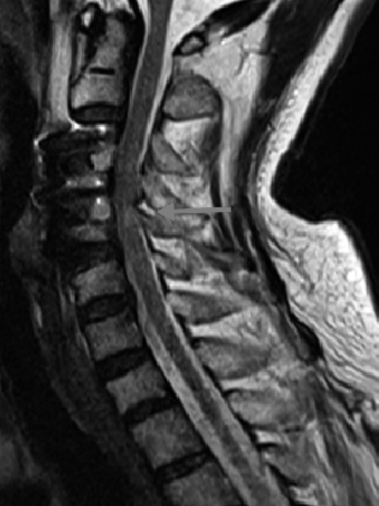
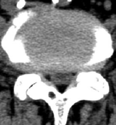
Based on these clinical and imaging findings, the patient underwent ACDF at the levels of C3-4 and C4-5 for cervical cord decompression. Neurological examination performed in the recovery room after surgery identified a grade 0/5 for the left ankle dorsiflexors and big toe extensor muscles, with no other change in strength identified. Immediate follow-up MR imaging revealed an increased thickness of the ligamentum flavum at the level of C4-5, with increased protrusion into the spinal canal, compared to pre-operative MR images (Fig. 2). A computed tomography scan of the lumbar spine was negative, ruling out acute lumbar disc herniation and lumbar spinal stenosis as possible causes of the loss of strength of the left ankle dorsiflexors and toe extensors (Fig. 3). Review of the operative procedures, including positioning of the lower limbs, did not reveal any probable cause of injury to the head of the fibula and the peroneal nerve. As well, the surgical procedure was reviewed from the intra-operative video, with no evidence of probable spinal cord trauma identified.
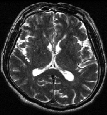
Based on these findings, thickening of the ligamentum flavum was determined to be the most likely cause of the left foot drop due to an acute anterior compression of the spinal cord at the level of C4-C5. This increased thickness of the ligamentum flavum was likely due to the reduction of the cervical lordosis after surgery. Therefore, a left side laminectomy and bilateral decompression were performed at the level of C3-C4, as well as at the level of C4-C5 to achieve a more complete decompression of the spinal canal. The neurological examination remained unchanged after the decompression surgery, with the motor impairments at the ankle and big toe persisting. On postoperative day 4, MR imaging of the brain was performed to rule out a possible cerebral infarction (stroke). The MR scans were negative, with no change in signal intensity on both T1-and T2-weighted images (Fig. 4). Strength of ankle dorsiflexors and of the big toe extensors recovered progressively, with a grade 3/5 assessed on the postoperative 7th day. Follow-up examination at 6-months, post-surgery, indicated full recovery of strength, to a grade of 5/5.
DISCUSSION
A foot drop can be reflective of a number of myopathies and neuropathies, ranging from direct trauma to the peroneal nerve to myotonic dystrophy and other types of distal muscular dystrophies, such as Welander, Nonaka and Laing types of distal myopathies [6]. Although peripheral and lumbar spine lesions are the most common causes of foot drop [6,11,15,18], lesions of the central nervous system and upper motor neurons can also result in a drop foot.
The motor pathways of the lower limbs are organized somatotopically, from the medial motor cortex, through the internal capsule and the ventral gray matter of the anterior horn cell at the spinal cord [18]. The axons of the L4 and L5 spinal nerves originate at the anterior horn of the spinal cord, with the nerve roots of these two peripheral nerves being in proximity at the lumbosacral trunk and the sacral plexus. The lateral trunk of the sciatic nerve becomes the common peroneal nerve, At this point, the common peroneal nerve divides into the superficial and deep peroneal nerves [15]. Because of its superficial position around the head and neck of the fibula, the peroneal nerve can easily be compressed by an external force, such as prolonged sitting with legs crossed. The peroneal nerve can also be directly damaged by fractures of the head and neck of the fibula or become entrapped in the fibular tunnel [6,11,15,18]. Common lumbar spine pathologies that can cause a foot drop include L4-L5 HNP and associated radiculopathy, foraminal stenosis or spinal canal stenosis. Other possible causes of peroneal neuropathy include metabolic changes, such as diabetes mellitus, drug toxicities, severe weight loss, inflammatory lower motor neuropathy or vascular pathology [1,11,15]. Central nervous system and upper motor neuron causes of foot drop have also been identified, including primary and metastatic brain tumor, ischemic stroke, intra-cranial hemorrhage, head trauma, cortical dysplasia, and abscess [1,5,8,9,13,16]. Although less common, a foot drop has also been documented in cases of incomplete traumatic spinal cord injury, thoracic disc herniation, cervical spondylosis, and spinal tumor [17].
In the case reported here, the foot drop was associated with a complete paralysis of the muscles of the anterior compartment of the lower leg (muscle grade of 0/5) and an increased thickening of the ligamentum flavum at C4-C5. This increased thickening after surgery most likely resulted from reduction of the cervical spine lordosis with surgery.
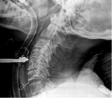
Thickening of the ligamentum flavum, with associated stenosis of the spinal canal at C3-4, C4-5, C5-6 and C6-7 levels was identified on the pre-operative dynamic MR imaging examination (Supplementary Fig. 1). Stenosis was also evident on intra-operative radiological examination, with the patient in a position of neck hyperextension (Fig. 5). Hyperextension of the neck during surgery likely did increase spinal stenosis, resulting in the anterior compression of the spinal cord and a foot drop.
Anatomically, the cervical spine has an intricate structure, including the myelinated axons of ascending (sensory) and descending (motor) tracts, and gray matter, a dense region containing neuron cell bodies and cell processes, along with their synapses, neuroglial cells and capillaries [3]. Compression of the cervical cord can damage some or all of these structures, resulting in a drop foot. Yamaura et al. [19] investigated the mechanisms of pathological changes of the spinal under conditions of chronic mechanical compression in a mouse model, reporting degeneration of the descending pathways of the anterior and lateral columns, as well as degeneration of the ascending pathways of the posterior column. Based on their findings, Yamaura et al. concluded that cell apoptosis due to chronic compression can produce destructive changes in both sensory and motor pathways in the spinal cord, resulting in irreversible neurologic deficit. Therefore, pre-existing spinal cord damage to motor and sensory pathways was likely to be present in our patient, owing to the long-standing mechanical compression of the spinal cord at C3-C4, C4-C5 and C5-C6 due to spinal stenosis, HNP, and thickened ligamentum flavum. The symptoms were exacerbated by intra-operative neck hyperextension.
ACDF is a safe and effective procedure. In a review of 1576 patients who underwent ACDF, Nanda et al. reported worsening of pre-operative symptoms in 14 patients (0.88%), including 5 patients with symptoms of myelopathy and 9 with symptoms of radiculopathy [12]. In their case series analysis of 83 patients who underwent ACDF, Song et al. reported dysphasia as a complication in 4.8% of cases [14]. Comparatively, in their case series of 57 patients, Lin et al. reported complications in 19.3% of cases, which included cerebrospinal fluid leakage, epidural hematoma, C5 radiculopathy, and dysphasia [10]. In a review of 11,817 patients who underwent spinal surgery, Cramer et al. identified only 21 (0.29%) patients with new onset of major neurological deficits immediately post-surgery, including a quadriparesis due to a spinal cord hematoma [2]. Overall, the most common complications of ACDF is dysphasia and worsening of pre-existing neurological symptoms.
In the case presented here, neck extension had been verified by the anesthesiologist before surgery, with no complaints reported by the patient and no abnormal neurological findings. Therefore, the surgeon could not pay attention to check neck hyperextension at the marking film (Fig. 5). Intra-operative neurophysiological monitoring of spinal cord activity may have been useful for early detection of spinal cord damage. This case does raise questions regarding the usefulness of somatosensory-evoked potentials alone as a monitoring tool for the detection of new onset major neurologic deficit during ACDF [2].
CONCLUSION
The case presented here describes a unique complication of ACDF, a left side foot drop, which resulted from an indirect injury to the spinal cord due to pre-existing risk factors, namely cervical spinal stenosis. Specifically, intra-operative hyperextension of the neck increased anterior compression on the spinal cord, despite any negative pre-operative assessment for risk factors. Therefore, in cases of acute onset of drop foot after ACDF, central causes should be investigated.