INTRODUCTION
Choi et al. [2] reported L5-S1 interlaminar approach to treat L4-5 down migrated disc herniation. Developed from this previous technique, authors present a technical case report for simultaneous L4-5 transforaminal and L5-S1 interlaminar PELD in L4/5 down migrated disc herniation.
CASE REPORT
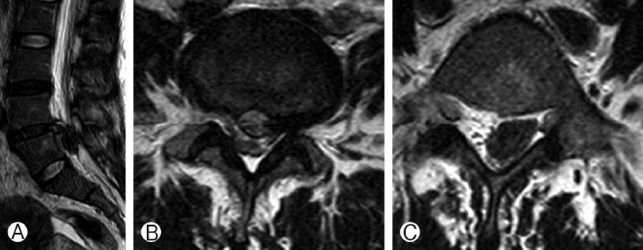
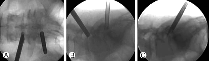
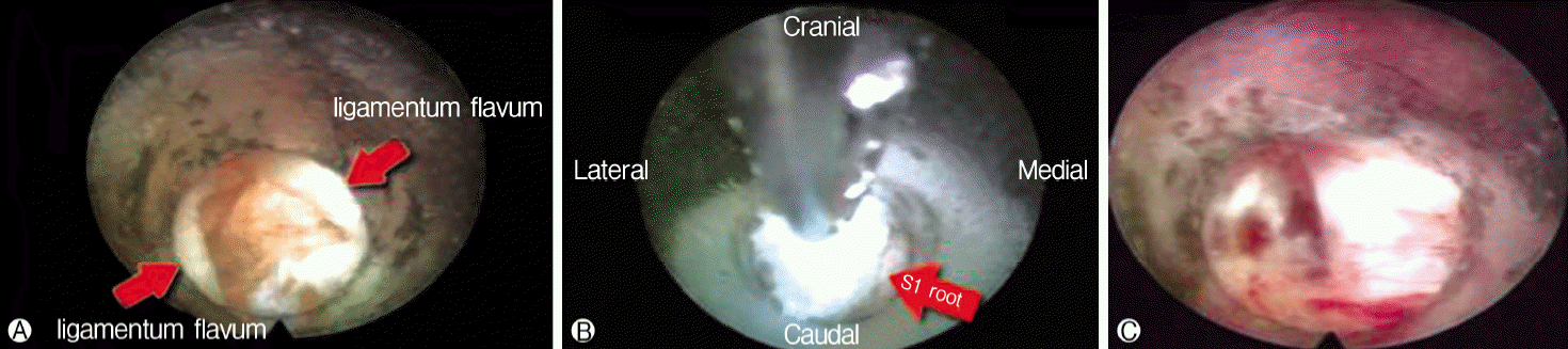
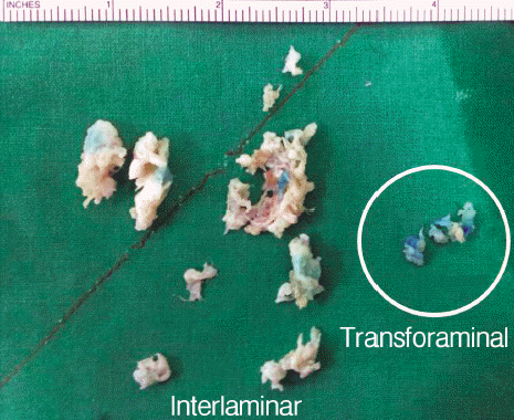
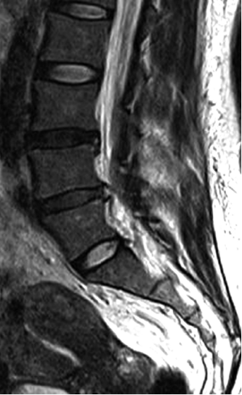
A 44 years-old woman presented with severe left lower extremity radiating pain and back pain, and ambulation was not possible. MRI showed left sided large amount of disc herniation with down migration at L4-5 (Fig. 1). The patient did not want open surgery nor general anesthesia. We decided for percutaneous endoscopic lumbar discectomy (PELD). Since significant amount of disc material is present in both subannular lesion and down migrated portion, transforaminal approach for L4-5 and interlaminar approach for L5-S1 were planned. Left side transforaminal approach at L4-5 was first performed to decompress subannular disc herniation (Fig. 2). Down migrated disc was also to be removed at transforaminal approach but forceps could not reach to grasp it. Rigid working channel was introduced through L5-S1 left interlaminar window. Down migrated herniated disc was seen just after splitting ligamentum flavum (Fig. 3). The multi-fragmented disc was removed completely (Fig. 4 and 5). Two 0.8 cm sized post-operative scars are observed. The pain disappeared and patient was discharged next day.
DISCUSSION
PELD is recognized as a minimal invasive surgical option for treatment of disc herniation. One of the biggest advantages of PELD is preservation of the soft and bony tissues that are usually resected in standard discectomy [4]. Other advantages include minimal anesthesia, shorter hospital stay, faster return to activities of daily living, lower estimated blood loss during surgery, lower cost of postoperative care, and lower infection rates [6]. Open lumbar discectomy is gold standard to treat herniated disc and it is especially true in difficult case like this. Since this is only case report, superiority of the technique could not be proved but authors want to report one minimal invasive technique to treat down migrated disc herniation.
For treatment of migrated disc herniation, numerous methods have been proposed. Lee et al. [5] treated migrated disc herniations by “half and half technique” and “epiduroscopic technique”. They reported that down migtrated below lower level pedicle showed unfavorable result and discourage PELD in those cases. Choi et al. [1] reported transforaminal foraminoplasty to approach for highly down migrated disc herniation. By removing part of facet joints, working channel can be tilted in steeper angle to reach down migrated disc. Kim et al. [3] reported transforaminal contralateral apparoch in highly down migrated disc to decrease chance of exiting nerve root damage. Choi et al. [2] reported treatment L4-5 down migrated disc herniation via L5-S1 interlaminar approach. Du et al. [7] reported good outcomes in treatment of down migrated disc via translaminar PELD.
Authors thought that it would be difficult to remove whole disc from only transforaminal or interlaminar approach. There would be insufficient decompression if only one approach was performed. Reason for not removing transforaminal working channel (Fig. 2) was that authors wanted to use triangulation technique as arthroscopic surgery which uses one portal as a viewing portal and the other as a working portal. However, disc fragments were well removed with each approach, and biportal manipulation was not necessary. Some would questions that L4-5 transforaminal approach was really necessary. Amount of sub-annular disc could not be ignored and could not be removed by interlaminar approach from L5-S1. From L4-5 transforaminal approach, complete removal of down migrated disc fragment could be expected in if forcep can grasp part of all herniated fragments. This is easy if there is only one fragment. However in this case, there were multiple fragments and could not be removed from transforaminal approach. Still, L4-5 transforaminal approach should precede interlaminar approach in L45 down migrated disc herniation, if surgeons think subannular disc is large and should be removed because surgery can be finished with only transforaminal approach.
Indication of this technique is limited to L4-5 down migrated disc with wide L5-S1 interlaminar window. This technique is similar from previous technical note for L4-5 down migrated disc herniation via L5-S1 interlaminar approach [2]. Choi et al. [2] did not define criteria for wide L5-S1 interlaminar window. Wide interlaminar window can be referred as upper roof of interlaminar space should be at least above the anatomical line bisecting the midpedicular level of L5 on the coronal image or AP X-ray. Diameter of working channel is about 8 mm. Surgeons can measure preoperatively in AP X-ray that interlaminar window of ipsilateral side can accommodate 8 mm diameter working channel. Once working channel is introduced in epidural space, working channel should be tilted caudally in maximum angle and approach to L4-5 down migrated disc is possible.