Realizable Minimally Invasive 1-Day Lumbar Interbody Fusion Surgery: No General Anesthesia, No Hemovac Insertion, No Skin Suture Surgery, and Early Ambulation
Article information
Abstract
Objective
The lumbar interbody fusion surgery, patients commonly have severe pain, requiring adequate bed rest for a long time. We performed a 1-day minimally invasive spine (MIS) lumbar interbody fusion that required no hemovac insertion and no skin suture and led to early ambulation. Here, we report the surgical procedure and results.
Methods
This study was designed as a retrospective review. From January 2013 to August 2014, 49 patients who received the MIS TLIF for 1-day MIS lumbar interbody fusion surgery were included in this study. The surgical procedures performed were as follows: (1) epidural catheter insertion; (2) midline subdermal dissection procedure; (3) MIS TLIF; (4) bleeding control procedure; (5) percutaneous transpedicular screwing; (6) tight subdermal plan suture; (7) skin sealing procedures. Postoperatively, wound dressing was not needed. Epidural catheter was removed on the second day after the operation.
Results
Average intraoperative bleeding was 128.6 mL per level. The average operation time was 78.9 min. per level. An average midline skin incision was 2.8 cm per level. The possible ambulation time was 0.94±0.88 day. The discharge time after antibiotic injection for 3 days was 4.88±1.51 days. In the corresponding order of preoperative and immediate postoperative, 3-month, 6-month, and final follow-up, Postoperative VAS (back), VAS (leg) and ODI improved significantly immediate postoperatively (p<0.0001). Postoperatively, there was no cases of revision due to hematoma.
Conclusion
The results indicated good clinical results of the 1-day minimally invasive lumbar interbody fusion surgery, without any serious complications.
INTRODUCTION
The incidence of degenerative spinal diseases that require lumbar interbody fusion surgery has increased with an increase in the elderly population. However, following lumbar interbody fusion surgery, patients commonly have severe pain and require long periods of adequate bed rest. Moreover, associated complications can occur, leading to a delay in rehabilitation.
An extensive body of research supports Minimally Invasive Surgical (MIS) techniques as effective in decreasing postsurgical morbidity and improving postoperative recovery [13].
However, because of the risk of complications, patients who are in need of fusion surgery, especially the elderly, tend to avoid surgery. Furthermore, shortening of the length of surgery and hospitalization is an important factor in the onset and recovery of complications in patients with a systemic disease.
In fact, a prior study has reported outcomes following 1-day fusion surgery [15]. However, in this study, although patients were discharged on the same day as surgery, it did not mean that care was also terminated. Rather, this study compared and verified that same-day discharge did not cause complications. In fact, stitch removal and patient care were managed in outpatient appointments after discharge. Thus, the patients were not truly discharged but were transferred to home care. The fact that the patients were still in need of stitch removal and hospital care signified that the patients were not completely discharged from the hospital.
Ambulation is a critical aspect of rehabilitation and positively affects patients’ recovery. In fact, the main reasons patients stay in hospital are as follows: (1) postoperative pain management, (2) management and removal of any drainage tubes, and (3) postoperative wound care. Thus, if these procedures were unnecessary, true 1-day fusion surgery would be possible.
We performed a 1-day MIS lumbar interbody fusion that did not require Hemovac insertion or postoperative sutures and allowed early ambulation. Here, we report the surgical procedure and results.
MATERIALS AND METHODS
This study was designed as a retrospective review of clinical and surgical parameters. From January 2013 to August 2014, 49 patients who underwent 1-day MIS transforaminal lumbar interbody fusion (TLIF) surgery were included in this study.
All patients underwent MIS TLIF using an MIS retractor system (Tubular/Caspar/Taylor) and MIS decompression technique (unilateral decompression/bilateral decompression/unilateral approach bilateral decompression). Two cases were being treated for foraminal stenosis, one for recurrent Herniated Nucleus Pulposus (HNP), 13 for spinal stenosis, and 33 for spondylolisthesis.
1. Patients
This study examined surgeries conducted between January 2103 and August 2014 by reviewing the medical charts of 49 patients. Patients with spinal stenosis, spondylolisthesis, foraminal stenosis, or recurrent HNP were included in this study, all of which were in need of decompression, as well as pedicle screw fixation and fusion.
2. Operative techniques
Decompression was performed using the basic MIS TLIF procedure as described below.
3. Technique for MIS TLIF
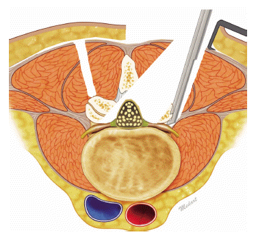
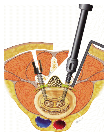
The MIS TLIF procedure was performed on the symptomatic side. C-arm guidance was used to determine the disc space and to draw the lateral pedicle line in the fluoroscopic anterior posterior view. After a vertical skin incision in line with the lateral pedicle. After a complete facetectomy, the ligamentum flavum was removed to expose the lateral border of the ipsilateral nerve root. The retractor was angled medially, The patient was tilted laterally to decompress the contralateral side. Extensive decompression was performed, which included decompression of the central stenosis and contralateral side [1,6,9]. A discectomy was also performed. A single, banana-shaped polyetheretherketone interbody cage filled with only autologous local bone was inserted. After interbody fusion, the retractor was removed, and the same procedure was repeated for each segment. Ipsilateral percutaneous pedicle screws were inserted through the same skin incision. Contralateral percutaneous pedicle screws were placed using a mirror incision under fluoroscopic guidance.
Additional surgical procedures performed were as follows:
(1) Epidural catheter insertion for anesthesia and postoperative pain control; this allowed the procedure to be performed without a general anesthesia and controls postoperative pain effectively such that patients can ambulate shortly after surgery.
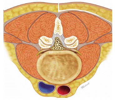
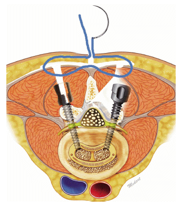
(2) midline subdermal dissection procedure: this procedure can reduce the size of the skin incision, and the tension of the skin can reduce the risk of postoperative hematoma. Although it is a midline incision, dissection is performed at the subdermal level, and TLIF, decompression are performed via the paraspinal plane. PLIF, which comes in contact with the midline structure, can induce central accumulation of blood and eventually cause a postoperative hematoma (Fig. 1)
In addition, advances in MIS techniques for TLIF have reduced the incidence of complications and morbidity associated with conventional TLIF [3].
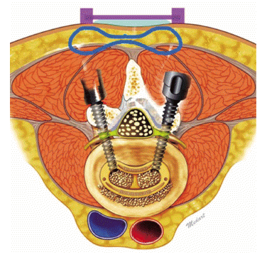
(3) MIS TLIF procedure (unilateral/bilateral) (Fig. 2).
(4) Percutaneous transpedicular screw insertion under the subdermal dissection plane (Fig. 3).
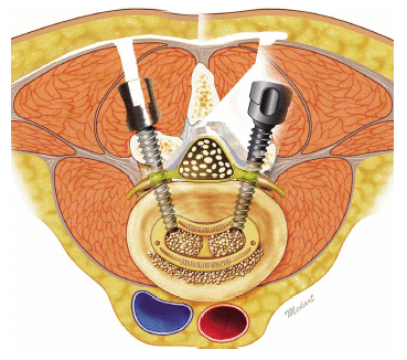
(5) Blood loss control procedure: limiting blood loss is the most crucial step, as hemostasis is a key aspect in 1-day fusion surgery. Our procedures included the following: (A) meticulous bleeding control; (B) fibrinogen/thrombin-based collagen fleece bleeding control; (C) fluid-type anti-adhesive agent, which can stop venous bleeding using hydrostatic pressure; and (D) GelfoamⓇ covering, which acts as a barrier that stops bleeding that occurred outside the spinal canal, i.e., from the muscle, from coming into the canal (Fig. 4).
(6) Tight subdermal plane suture (conjoined suture of split fascia and subdermal skin) (Fig. 5).
(7) Skin sealing procedures: secure skin and zip surgical skin closure systems (Fig. 6).
A postoperative wound dressing was not needed. The wounds were checked every 3-4 days. The epidural catheter was removed on the second day after the operation. Intravenous antibiotics were administered for 3 days after the operation.
Epidural catheter insertion enables additional pain control. Even if an IV PCA is used, patients experience the most severe pain during the first two days post-operatively, which can be controlled continuously via the epidural catheter.
RESULTS
We examined surgery-related results, the intraoperative and postoperative conditions, postoperative complications, and clinical results by using the Visual Analogue Scale (VAS) and Oswestry Disability Index (ODI) immediately (1-2 days), and 1 month, 3 months, 6 months, and 12 months postoperatively.
1. Demographics
The mean age was 65.27±9.57 years, and the sex ratio was 20:29 (male:female). The average follow-up period was 26.04±7.25 months.
Regarding the number of segments involved in the operation, 33 patients underwent one segment; 13 patients, 2 segment; and 3 three patients, 3 segment operation (average: 1.39±0.61 segment).
2. Clinical outcomes
Average intraoperative bleeding was 178.47±73.70 mL (per level: 128.60 mL). The average operation time was 109.49±32.71 min (per level: 78.90 min). Average midline skin incision length was 3.90±1.18 cm (per level: 2.80 cm).
The possible ambulation time was 0.94±0.88 day. The discharge time after 3 days’ antibiotic administration was 4.88±1.51 days.
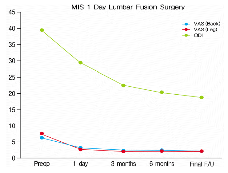
The VAS (back) were as follows: 6.33±0.94, 3.14±1.12, 2.47±0.58, 2.29±0.65, and 2.31±0.77; VAS (leg): 7.37±0.70, 2.69±0.85, 2.29±0.46, 2.14±0.58, and 2.24±0.80; and ODI: 39.37±3.05, 29.29±5.78, 22.59±2.99, 20.27±2.59, and 18.63±3.13 for the preoperative, immediate postoperative, and 3-month, 6-month, and 12-month follow-up values, respectively. Postoperative VAS (back), VAS (leg), and ODI improved significantly immediately postsurgery (p<0.0001) (Table 1).
In terms of postoperative complications, there were two cases of transient motor weakness (both cases recovered sufficiently after the follow-up period), four requiring wound suture due to avulsion of the surgical field (all cases healed completely after the follow-up period), one of dural tear, and two of cage subsidence or implant failure. No cases required revision due to hematoma.
3. CASE
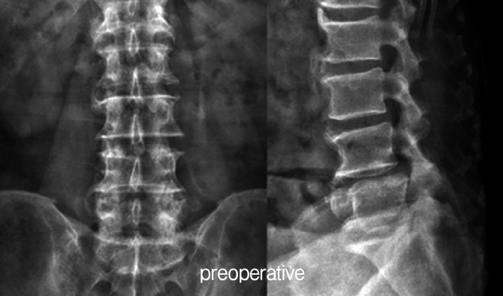
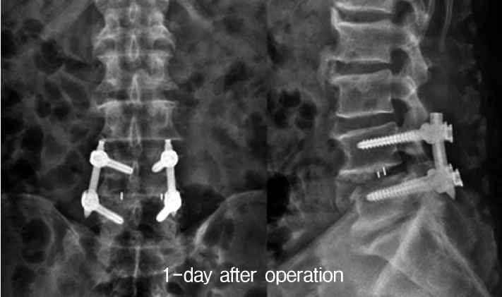
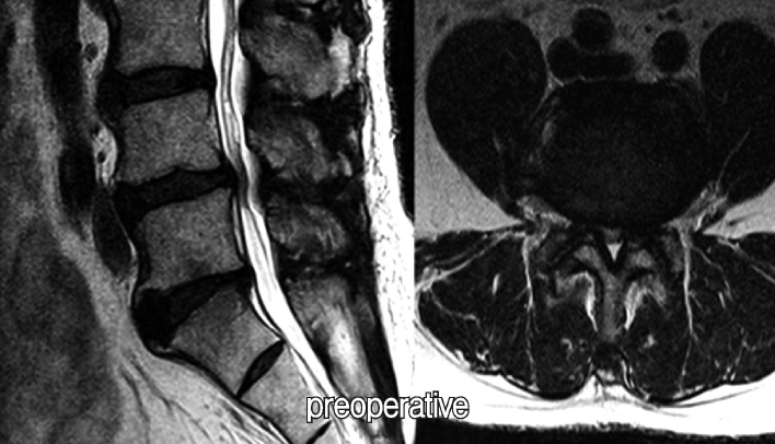
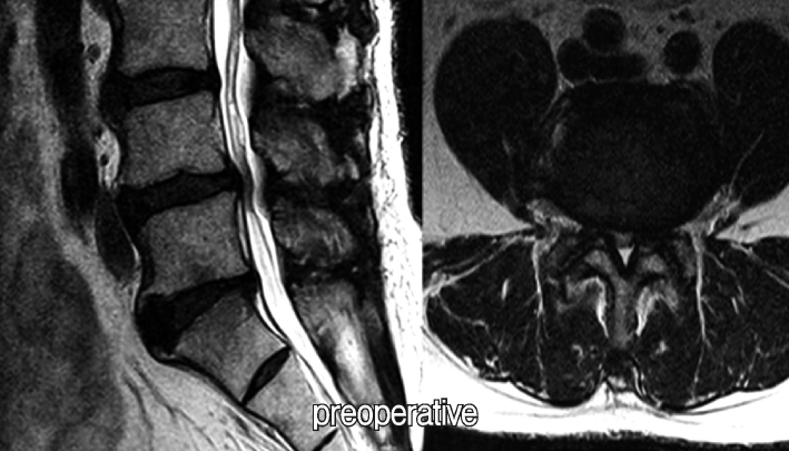
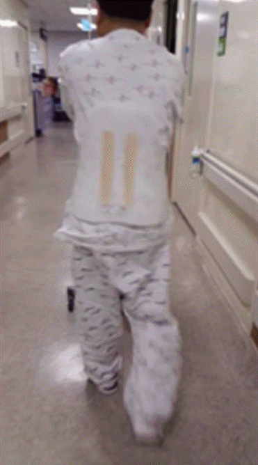
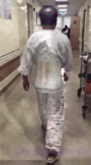
56-year-old male who visited the hospital for severe, radiating low back pain and neurogenic claudication present for more than a few months. The 1-day postoperative MRI showed sufficient decompression with MIS TLIF. The patient was capable of walking by the afternoon on the day of surgery. He was capable of discharge after 1 postoperative day (Fig. 7-12).
DISCUSSION
McGirt et al. [8] reported that with 2-level transforaminal lumbar interbody fusion, mean costs associated with perioperative surgical-site infection were significantly lower with MIS than with open procedures. Similar cost-savings have been reported [7,14,16,17]. The patients’ clinical outcomes were assessed via VAS and ODI, but fusion rate was not included in this study. The reason was that the present study sought to examine the viability of 1-day fusion surgery, and fusion rate was intentionally ignored as the objective of this study was to describe the 1-day fusion surgery. However, considering the fact that MIS TLIF was performed for fusion, when compared with conventional TLIF, MIS TLIF appears to achieve similar fusion rates, while reducing blood loss, soft tissue and muscle trauma, postoperative pain, and increasing the speed of recovery [9,11,12]. It would rather be meaningful to study the fusion rate in the long-term along with other parameters. An epidural catheter was inserted to control postoperative pain. While reducing the pain, it also reduces sensory functions, which may hinder the patient from recognizing postoperative hematoma-induced pain until neurologic deficits, such as motor weakness or cauda equina syndrome, occur. The appropriate dose should be calculated for anesthesia via epidural catheter to ensure that only postoperative pain is reduced.
This study has a limitation regarding patient discharge. In Korea, the government pays >60% of all medical costs through the National Health Insurance Service, therefore most patients are discharged after the sutures are removed. The absence of a significant difference in hospital stay between MIS TLIF and the 1-day fusion technique is due to the unique medical system of Korea. To compare cost effectiveness between MIS TLIF and the 1-day fusion technique. Hence, although this study has described a surgical technique that enabled real 1-day discharge, various assessments, such as ambulation, should be emphasized rather than the actual date of discharge [2]. Early ambulation can lead to early rehabilitation, which in turn leads to early recovery; ultimately, postoperative complications are minimized, leading to effective rehabilitation. National Health Insurance systems differ for each country, so there would be practical differences, but minimally invasive procedures should be studied to develop such surgical techniques.
Many physicians consider GelformⓇ as a foreign body and worry about mass effect and infection from leaving Gelfoam at the laminectomy site. Although there were no postoperative infections in the present study, the possibility of other issues that may be revealed in a larger number of cases should be noted.
Furthermore, this study did not distinguish patients using anticoagulants, thus did not develop a separate protocol; this should be addressed in the future as well. In addition, preoperative assessment items for 1-day fusion surgery should also be elaborated.
CONCLUSION
The results indicated good clinical results for the 1-day minimally invasive lumbar interbody fusion surgery without any serious complications. With the development of an effective infection control system for the lumbar interbody fusion surgery, an effective true 1-day lumbar interbody fusion surgery will be possible.
