INTRODUCTION
Microsurgical discectomy, which was introduced in the 1980’s, is now established as the standard surgical treatment for lumbar disc herniation (LDH). During microsurgical discectomy, the classic surgical technique for bone work is partial hemilaminectomy with or without medial facetectomy from the inferior surface of lamina (the spinolaminar junction), though the extent of laminectomy required depends on target fragment location and disc level.
For upper LDH (L1-2 or L2-3) treated using the classic laminectomy technique, wide laminectomy, including pars interarticularis or facet joint, may result in iatrogenic spondylolysis or segmental instability because of the anatomical characteristics of the upper lumbar spine. Recently, to prevent the risk of iatrogenic instability during surgery for upper LDH, we performed key-hole laminotomy (microsurgical translaminar approach), which has been used for foraminal disc herniation in lower LDH since 1998 [3]. Here, we review of our experiences from the clinical, radiological, and surgical perspectives.
MATERIALS AND METHODS
1. Indication and patient population
The indications for discectomy surgery were as follows: (1) Persistent severe low back pain (LBP) and radiating leg pain (LP) despite adequate conservative treatment; (2) Severe LBP and leg pain making daily life impossible; or (3) Severe paresis (motor grade 3 or less).
From January 2007 to December 2014, 28 patients with upper LDH underwent single level unilateral discectomy using a microsurgical translaminar approach. Eight patients were excluded from the present study due to short-term follow-up (<1 year) or follow-up loss. The remaining 20 patients were selected for this study. All surgical procedures were performed by two surgeons (S.G. Lee and S. Son).
2. Operative technique
After induction of general anesthesia, the patient is placed in the prone position. A midline 2-3 cm skin incision is made at the fluoroscopically marked level and subcutaneous tissue is sharply divided. After incising muscular fascia and sweeping paravertebral muscles laterally from the lamina, a Caspar-type retractor is introduced to expose the hemilamina of the upper vertebra and inter-laminar ligamentum flavum.
Under microscopic view, a round or oval, 6-8 mm sized fenestration is drilled through the lamina craniomedially to the facet joint. The location of the hole depends on the location and morphology of the targeted extruded disc or migrated disc fragment. After bone work, in most cases, the ligamentum flavum is reached as the next layer, but in a few cases, the epidural space is encountered immediately. Therefore, to prevent tearing of dura mater, we use a diamond drill during drilling the basal portion of the fenestration close to the epidural space. The ligament is removed carefully from the undersurface of the lamina with a 1- or 2-mm Kerrison punch, and then the spinal canal is entered to identify the traversing root. Because of the anatomical characteristics of the upper lumbar level, this procedure permits exploration of the intervertebral disc space and for migrated extruded fragments. Evacuation of the intervertebral disc is dependent on pre-operative planning and/or intra-operative findings. Figure 1 shows a 3-dimensional computed tomography (3D-CT) image demonstrating the extent of fenestration in a patient with preoperative upper LDH.
3. Outcome evaluation
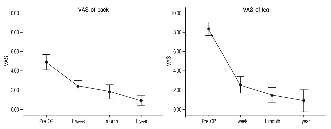
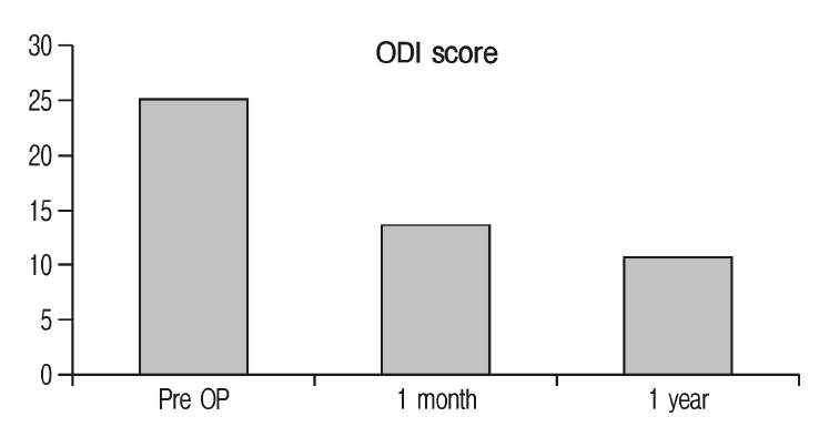
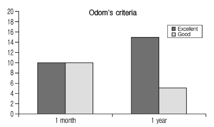
Clinical outcomes were assessed preoperatively and at each follow-up visit (1 week, 1 month, and 1 year postoperatively) using a visual analog scale (VAS) of the back and leg, Odom’s criteria, and Oswestry Disability Index (ODI) of quality of life.
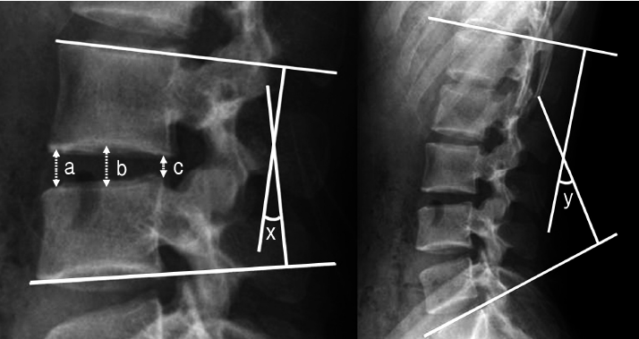
Lumbar MRI (magnetic resonance imaging), lumbar CT, and dynamic radiography were performed prior to surgery and lumbar MRI and CT immediately after surgery. In addition, dynamic radiography was performed at each follow-up visit. Disc height at index level, segmental range of motion (ROM), and total lumbar lordotic angle (Cobb’s angle) were used to evaluate radiologic outcomes (Fig. 2).
Perioperative outcomes were evaluated by investigating operating time, estimated blood losses (EBL), hospital stay, returntowork time, failure rate, recurrence rate, and surgical complications, such as, neurologic deterioration and wound infection.
4. Statistical analysis
Data management and statistical analysis were performed using the SPSS version 19.0 (SPSS Inc., Chicago, IL, USA). Results are expressed as mean values±standard deviations (SDs). The paired t-test was used to compare pre- and postoperative para- meters. Statistical significance was accepted for p-values of <0.05.
RESULTS
1. Demographic data and baseline characteristics
Among the 20 patients, there were 12 men and 8 women (mean age, 58.0±12.0 years; range 39-78 years). Surgery levels were L1-2 in 4 and L2-3 in 16 patients, and migration occurred in 14 patients. Average symptom duration was 2.4±1.7 weeks and weakness was accompanied in 8 patients. Demographic data and baseline characteristics including preoperative MRI characteristics and intraoperative findings are summarized in Table 1.
2. Clinical outcomes
Mean preoperative VAS of back was 4.9±1.1, and this decreased to 2.4±0.8 at 1 week to 1.8±1.0 at 1 month and to 0.9±0.7 at 1 year (p<0.001, paired t-test). Mean preoperative VAS of leg was 8.3±0.9, and this decreased to 2.5±1.2 at 1 week, to 1.5±1.1 at 1 month, and 0.9±1.6 at 1 year (p<0.001, paired t-test) (Fig. 3).
3. Radiological outcomes
Mean disc height at surgery levels was significantly decreased from 8.9±1.9 mm preoperatively to 8.2±2.3 mm at 1 year (p=0.043, paired t-test). Mean segmental ROM at surgery levels decreased from 6.4±2.6° to 3.0±1.8° at l year, but this was not significant (p=0.134, paired t-test). Degrees of spondylolisthesis preoperatively and 1-year were no different. However, mean total lumbar lordotic angle significantly increased from 26.8±10.8° to 36.6±10.6° (p=0.021, paired t-test). Radiological data are summarized in Table 2.
4. Perioperative outcomes
Mean operating time was 83.9±22.6 minutes, mean estimated blood loss was 75.6±55.5 mL, mean hospital stay was 8.6±3.8 days, and mean time to return-to-work was 20.3±5.80 days. No major surgical complication, such as, dura tear, neurologic aggravation, or wound infection, was encountered, and no perioperative morbidity related to general anesthesia, such as, a cardiopulmonary problem or deep vein thrombosis, occurred.
Target fragments were completely removed in all patients except one, who underwent nerve block at 2-weeks after surgery due to residual leg pain. There was one case of recurrence at 2 months after surgery, and the symptom was controlled using nerve root block. No patient required reoperation during the 1-year follow-up period.
DISCUSSION
During microsurgical discectomy for LDH, the extent of laminec tomy depends on target locations, which can be classified as disc, infrapedicle, pedicle, or suprapedicle levels longitudinally, and as central canal, subarticular, foraminal, or extraforaminal along the horizontal axis [14].
If ruptured disc material is located at the disc level on the subarticular side, even a small amount of rostral laminectomy will allow the target to be reached. However, if the ruptured disc material is located at the pedicular level and foraminal side (called “the hidden zone” by Macnab in 1971 [5]), wide rostral laminectomy, including isthmus, and a wide lateral laminectomy, including the facet joint, will ensure the target. However, in this case, wide laminectomy, including pars interarticularis and facet joint, may induce iatrogenic spondylosis or segmental instability. To overcome this risk, Di Lorenzo (1988) [3] introduced the “microsurgical translaminar approach” laminotomy technique for foraminal LDH, and over the years several reports demonstrated the safety and usability of this approach for “hidden zone” surgery in cases of lower LDH [1,6-8,10,12,13].
On the other hand, the extent of the laminectomy depends on spinal level. For example, for lower LDH, the rostral range of laminectomy can be minimized because the disc space lies approximately at the level of the interlaminar space [4]. Also, the lateral range of laminectomy (i.e., medial facet joint violation) can be minimized because the lateral border of the traversing root is contained to lamina width [16]. However, as the spinal level progresses cephalad, the rostral range of laminectomy to reach the disc space should be extended to the isthmus level because the disc space becomes more cephalad in relation to the interlaminar space [4]. Also, the lateral range of laminectomy should be extended to the facet joint or lateral border of isthmus because the lateral border of the traversing root is beyond the medial margin of the facet joint and the width of isthmus narrows [11,16]. This raises an important point for surgeons to consider, that is, in upper LDH, extended rostral and lateral laminectomy can reach the isthmus and facet joint and induce iatrogenic spondylolysis and segmental instability. Thus, to prevent iatrogenic spondylolysis in cases of upper LDH, the range of laminectomy should be carefully limited.
According to Reulen [11] and Ebraheim [4], lamina height increases with lumbar spinal level and the distance between the inferomedial-most aspect of the inferior facet and disc space decreases. Interestingly, based on these anatomical characteristics of the upper lumbar spine, a 6-8 mm sized translaminar fenestration is enough to ensure access to migrated disc fragments as well as disc space. This “key-hole laminotomy” technique, can prevent amputation of the isthmus and complete violation of the facet joint during discectomy in cases of upper LDH.
Regarding clinical outcomes, in the present study, improvements in back and leg VAS scores, ODI scores, and patient satisfactions according to Odom’s criteria were all favorable. On the other hand, from the radiological aspect, although disc height decreased significantly, segmental ROM and degree of spondylolisthesis were unaltered. In addition, total lumbar lordosis was significantly improved. These findings mean iatrogenic segmental instability can be prevented via key-hole laminotomy in cases of upper LDH.
Mean operating time and EBL were reasonable enough, as compared with those mentioned in a previous report on lumbar microsurgical discectomy [2], [15]. Also, surgical outcomes, including complication, failure (only 1 of the 20 patients required postoperative nerve root block for residual leg pain), and recurrence (none) rates were favorable.
Despite our favorable findings, this study has several limitations. In particular, the number of patients was too small and the follow-up period too short to allow generalizations of results. Additional study is required to compare the described key-hole laminotomy technique with conventional microsurgical discectomy in patients with upper LDH.
CONCLUSION
Despite its small cohort and short follow-up, the present study demonstrates that key-hole laminotomy (the microsurgical translaminar approach) is useful for preventing iatrogenic spondylolysis and segmental instability in cases of upper LDH. We suggest key-hole laminotomy be considered as a safe surgical option for treating upper LDH.


