INTRODUCTION
For the past decades, pedicle screw fixation has been regarded as a standard measure to obtain a rigid internal stabilization of the unstable spine. This system has been used successfully in various degenerative spinal disorders, including spondylolisthesis and stenosis because it has the advantages of providing three-column fixation, facilitating the instrumentation of short segments, and maintaining the anatomic or desired sagittal alignment of spinal curvature. In order to achieve these merits, a firm anchorage of the screws to the vertebral bodies is required and this is directly related to the quality of bone into which screws are placed [1,2,3]. Otherwise, the major complications of instrumentation such as loss of fixation or loosening of the construct might occur, especially in osteoporotic vertebrae.
Maintaining a constant screw construct would not be a simple issue after fusion surgery for the patients with osteoporosis and lots of different methods to improve the purchase of screws has been investigated using animal or cadaveric model. These include the augmentation of the screws with polymethylmethacrylate [4,5], modification of the design of the thread, the shape, and surface of the screws [5,6]. Despite these devise, there were no definite description of the benefit of these augmentation in maintaining the screw construct over a certain period of time after its performance to the patients.
In the current study, we performed the fusion surgery to the patients with degenerative spondylolisthesis with screw augmentation using the hydroxyapatite and compared its result to that of those without augmentation. This hydroxyapatite has already been reported to have the advantage of being resorbed and replaced during healing and normal bone remodeling [7,8]. Because the real parameters for measuring the screw purchase strength such as extraction torque or pull-out strength cannot be measured from the actual patients, several indirect radiographic parameters were measured to assess the maintenance of the screw construct during the each follow-up. This comparative study might provide the tip of suitability of hydroxyapatite cement for augmenting the initial fixation of screws and its advantage in maintaining a constant screw construct in senile, osteoporotic vertebrae.
MATERIALS AND METHODS
1. Patients
The 21 consecutive patients who were diagnosed as grade I degenerative spondylolisthesis on L4-5 level with osteoporosis and then treated with transforaminal lumbar interbody fusion (TLIF) operations between December 2002 and June 2004 at our institution with the minimum follow-up period of two years were included in this study. The diagnosis of osteoporosis was based on the measurement of bone mineral density (BMD) by T-score of less than -3.0 by dual-energy X-ray absorptiometry (DEXA) performed both on the femoral joint and lumbar vertebral body before operation. Among these, nine patients were treated by TLIF with screw augmentation with hydroxyapatite and the rest 12 patients were treated with TLIF only. The choice of the surgical modality was arbitrary according to the consent from the patient to pay for the extra cost for using the hydroxyapatite cement after they were explained as to the possible benefit that could be yielded by performing the screw augmentation. The augmentation group consisted of eight female and one male patient with the mean age of 66.6 years (range; 57 to 73 years) and the all twelve patients in the TLIF only group were female with the mean age of 66.9 years (range; 62 to 77 years). The mean T-scores after BMD on the lumbar vertebrae and femoral joint were -3.45 and -2.7 for the augmentation group and -3.54 and -2.7 for the TLIF only group. The detailed informations on the demographic features of both groups are listed in Table 1.
2. Insertion of pedicle screw
In all 21 patients regardless of the group, the TLIF operations were performed by the same senior surgeon (H.Y. Lee) in the author list. After the performance of lumbar interbody fusion using the polyethyetherketone (PEEK) cages, standard anatomical landmarks from intraoperative fluoroscopy were used for the identification of the pedicles and the confirmation of the proper position of the screws. The entry point to base of pedicle was passed by electric drill with 4 mm-diameter burr instead of using standard awl or tapering tool to avoid injury to the cancellous portion of the pedicle even before screw insertion. Then the screw path in the body was created with 10 gauze Poverty needle (Kyeongwon Medical, Seoul, South Korea) for vertebroplasty, which has the property of outer diameter of 3 mm and inner diameter of 2.2 mm. In the nine patients who agreed with the use of hydroxyapatite cement, needle was drawn back about 1 cm to create a void for all pedicles. About 2 cc of BoneSource hydroxyapatite cement (Stryker Howmedica Osteonics, Kalamazoo, Mississipi) was injected through the needle. Due to the mud-like nature of hydroxyapatite cement, tapping of inlet into the needle was necessary to achieve a complete injection. The typical mixture of these BoneSource cement consisted of liquid to powder ratio of 5 cc to 10 gm. We usually injected the BoneSource cement just after mixing without haste because unlike PMMA cement, pedicle screws could be inserted even after full hardening of hydroxyapatite cement. Then, bilaterally for each vertebra, a 6.5 mm diameter and 45±5 mm long Diapason System Pedicle Screws (Stryker Osteonics, Allendale, New Jersey) were inserted into the pedicles. The cement was allowed to harden for 24 hours after implantation with the patient in bed rest.
3. Radiological assessment
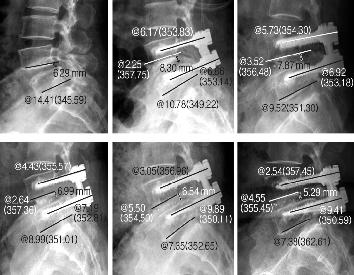
The plain antero-posterior (AP) and true lateral radiographs were checked serially during operation, immediately after the operation, 1 month, 3 months, 6 months, 1 year, and 2 years after the operation. The absolute values of the angles between the vertebral bodies at the operated level, between the upper and lower screws, between the inserted screw and lower endplate of corresponding vertebral body, and posterior disc height changes were serially measured from the lateral radiographs of the each corresponding period. The mid-axial line of each screw was used for the angle measurement. Because the two screws inserted for each vertebral body usually were not exactly parallel to each other on plain radiography, the screws inserted on the side of laminectomy and fascetectomy during TLIF were chosen for the radiographic measurement. These radiological changes were measured with aid of the PiView program (Infinitt Co., Seoul, South Korea) and the mean values were recorded after twice respective measurement by the first author (J.H. Lee) who was not involved with surgery. For the comparison between the two groups, (1) the absolute values of the mean angle and height at each corresponding period, (2) setting the measured values during the operation as index point, the amount of changes in angles or height over the time from index point to the each period, (3) the amount of changes in angles or height between each serial period were calculated (Figs. 1 and 2).
4. Statistical analysis
An analysis of variance (ANOVA) was conducted to assess the significance of the differences among the values measured at each follow-up period in each group, while an independent two sample t-test and a Wilcoxon rank sum test were performed to evaluate the difference of the amount of changes that have occurred during each sequential time-interval and from index point to each period on follow-up.
RESULTS
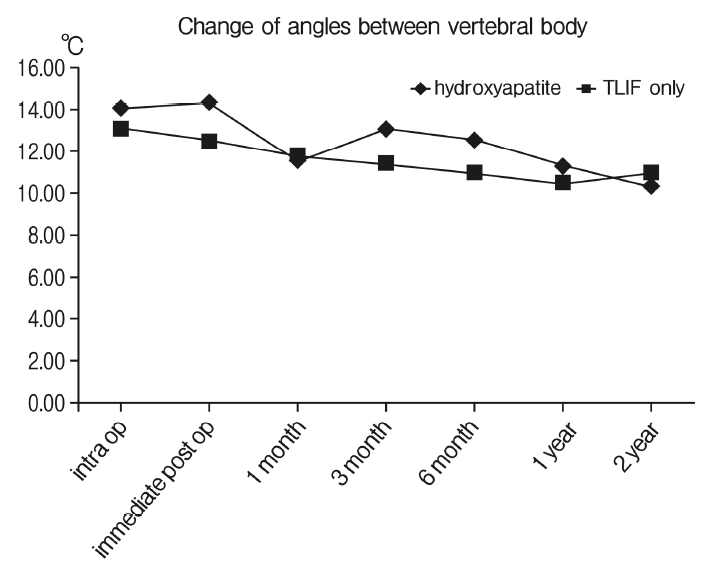
1. Angle changes between the vertebral bodies
The mean angles between the vertebral bodies at the operated level have reduced over two years of follow up from 14.09° to 10.3° for the hydroxyapatite augmentation group and 13.05° to 10.84° for the TLIF only group (p>0.05). Of all the comparison parameters between the two groups, only the mean difference of the degree of angle reduction occurred during the period from the operation to post-operative 6 months between the two groups showed statistical significance (-0.93° for the former vs -2.9° for the latter group, p<0.05). The results of the angle changes between the vertebral bodies for each group are summarized in Table 2 and Figure 3.
2. Changes of the posterior disc heights
The mean posterior disc heights between the vertebral body at the operated level have significantly reduced for each group over two years of postoperative period from 9.48 mm to 5.39 mm for the augmentation (p<0.05) and from 10.08 mm to 6.61 mm for the TLIF only group (p<0.05). However, the amount of reduction occurred between each period or from the index point to certain postoperative period showed no significant difference between two groups. The results of the changes in the posterior disc heights for each group are summarized in Figure 4.
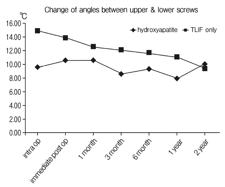
3. Angle changes between the upper and lower screws
Although statistically not significant, the mean angle between the upper and lower screws showed gradual decrease from 14.86° to 9.42° for the TLIF only group while well maintained for the augmentation group from 9.62° to 9.95°. However, the degree of angle changes occurred between each period or from the index point to certain postoperative period showed no significant difference between two groups. The results of the angle changes between the upper and lower screws for each group are summarized in Table 3 and Figure 5.
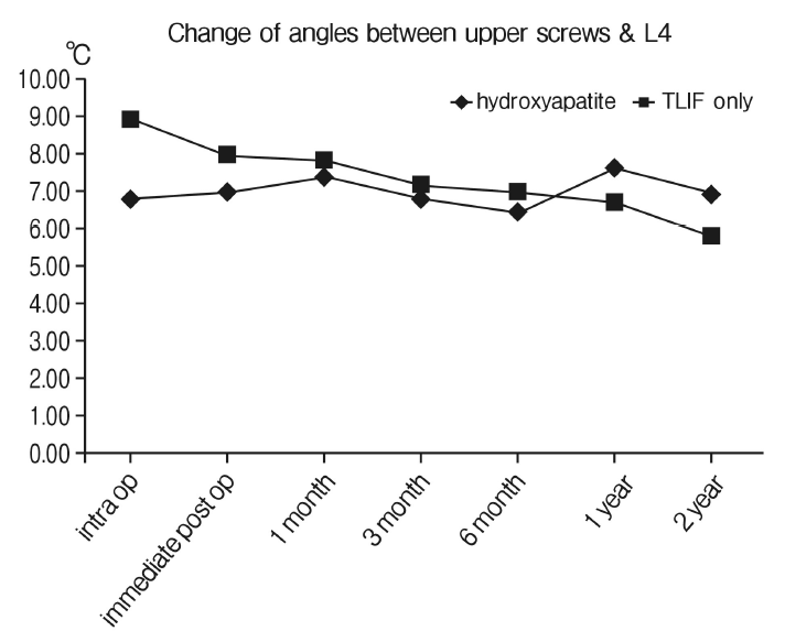
4. Changes of the angles between the upper screws and lower endplates of L4
Statistically significant degree of mean angle change of 1.17° between the upper screws and lower endplates of L4 occurred for the TLIF only group compared to 0.37° for the augmentation group during the period between the index point and immediate postoperative period (p=0.048). More significant angle changes have occurred for the TLIF only group over the period between the index point and postoperative 6 month (3.17° for the TLIF only group vs 0.72° for the augmentation group, p=0.003). Most of these significant angle changes seemed to have occurred for the TLIF only group between 1 month and 6 months after the operation as the change between 1 month and 3 months for the TLIF only was 1.53° compared 0.78° of the augmentation (p=0.02) and the change between 3 months and 6 months was 1.42° for the TLIF only compared to 0.21° for the augmentation group (p=0.001). Although it revealed that the mean angle change showed gradual decrease from 8.93° to 5.79° for the TLIF only group while well maintained for the augmentation group from 6.84° to 6.93°, this difference was of no statistical significance (p= 0.053). The results of the angle changes between the upper screws and lower endplates of L4 for each group are summarized in Table 4 and Figure 6.
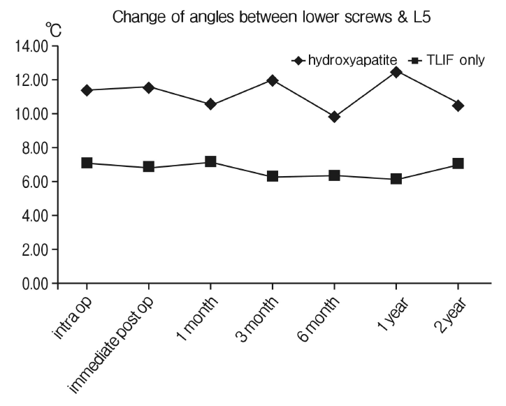
5. Changes of the angles between the lower screws and lower endplates of L5
Compared to the changes in angle formed between the upper screws and lower endplates of L4, the angles formed between the lower screws and lower endplates of L5 were relatively well maintained in both groups as the changes occurred only minimally from 11.3° to 10.41° for the augmentation group and from 7.02° to 6.99° for the TLIF only group (p>0.05). Consequently, the degree of angle changes occurred between each period or from the index point to certain postoperative period showed no significant difference between two groups. The results of the angle changes between the lower screws and lower endplates of L5 for each group are summarized in Table 5 and Figure 7.
DISCUSSION
Rigid fixation systems place high requirements on the load-carrying capacity of the screw-bone interface. Poor bone quality decreases the integrity of this surface, increasing screw micromotion and reducing stability and possibly fusion success [1]. In severely osteoporotic bone, pedicle screw use without supplementary augmentation might be precluded completely. Thus, a need exists for a measure to augment and strengthen the bone-screw interface that will reduce the risk of screw loosening and increase the endurance limit for pedicle screw fixation.
With this regard, the polymethyl methacrylate (PMMA) has been used in numerous animal or cadaveric studies in order to improve this implant fixation. The injection of PMMA was found to be effective augmentation method, increasing the pullout strength to approximately 150% of the initial pullout strength [9] and dramatically improve the resistance to screw toggle caused by cyclic caudocephalad loading [5]. However, although PMMA has been shown to significantly increase the pullout strength of pedicle screw fixation when inserted into the osteoporotic bones, there are several disadvantageous properties associated with the use of PMMA. These include the risk of tissue injury due to high exothermic polymerization temperature of 40 to 100°C [10], poor tolerance to fatigue, and its permanence in the body [3]. Moreover, the pedicle screws augmented with PMMA are also very difficult to remove, greatly complicating revision surgery [11]. Thus a biodegradable cement that has similar properties to PMMA, but that does not interfere with the natural bone healing or remodeling process, would be an attractive alternative for use in the augmentation of pedicle screws.
More recently, the suitability of nonexothermic, bioactive hydroxyapatite (HA) cements for pedicle screw augmentation has been investigated in this regard. Like PMMA, these cements can be injected or packed into irregular bony defects in the mixed, paste form. The injectable and in situ setting nature of this cement means that the cement-bone interface area is large which additionally contributes to the screw stability [3,7]. Also, the HA cements are biocompatible and bioresorbable, meaning that they are initially osteotransductive with rapid ossointegration after implantation and later osteoconductive with promoting new bone replacement directly on the cement surface without intervening fibrous tissue [8,12], thereby forming a strong bond between bone and implant while allowing for simpler revision in the long-term [13]. They solidify in minutes, and within four hours they attain 90% of their ultimate compressive strength, which is known to be as high as 60 Mpa in 24-hour wet compressive strength [14]. Moreover, unlike the polymerization of PMMA, the solidification of HA cement involves precipitation and is essentially nonexothermic [7], thereby precludes the potential for thermal necrosis of adjacent tissue.
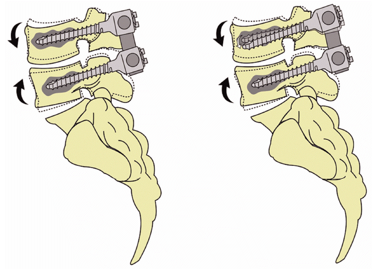
The result from the current study demonstrated that the statistically significant degree of mean angle change between the upper screws and lower endplates of L4 occurred for the TLIF only group compared to the augmentation group especially during the period between the index point and postoperative 6 month (Fig. 8). Meanwhile, the angles formed between the lower screws and lower endplates of L5 were relatively well maintained in both groups. There are no proven results reported previously in the literature to interpret this phenomenon but our assumption would be that the upper level of the construct is more vulnerable to the cephalocaudad mechanical loadings that are being transferred to the screw construct during the daily activity especially before acquisition of solid bony fusion between two segments. This seems natural in mechanical point of view, as most of the cephalocaudad loading during the daily motion would be initially transferred to the upper body and screw and then to the interbody cage during the stage of one to six months after TLIF as the non-fused segments during this postoperative period would not be able to act as a single unit. As the previous series investigating the effects of the posterior spinal instrumentation on the adjacent motion segments, the instrumented constructs produce higher segmental displacement values at the upper residual intact motion segment when compared with those of the intact spine, suggesting that more motion and loadings are being transferred to the upper level of the construct rather than lower level after fusion [15]. A more discrete biomechanical study such as the finite element analysis for the fused segments should be ensued to support this hypothesis.
In our surgical method, about 2 cc of the solution of the HA cement powder mixed with sodium phosphate was injected through the each tapped hole using the needle typically used for vertebroplasty before inserting the actual screws. This is somewhat different from the method of coating the pedicular screws with plasma-sprayed HA that is frequently used in the previous literature [16-18], as the augmentation would only involve the central vertebral body and tip of the each screw rather than the pedicle or entire length of the screw. It is well known that the stiffness of the pedicle provides most of the stability, contributing 80% to cephalocaudad stiffness [19], with stiffness of the vertebral body of secondary importance [9,20]. Moreover, reinforcing the entire screw-bone interface with cement has the advantage of increasing the compressive strength of the interface and it also increases the surface area of this interface [3], thereby decreasing the stress level. However, the result from the study by Renner et al. using the cadaveric model demonstrated that the average pull-out strength for the screws after injection of HA cement only to the distal tip of the screw hole was 1617N, which was not significantly different from 1852N measured after injection of HA cement along the entire length of the screw hole [21]. Since the initial stage of the screw loosening usually starts with the development of pendular motion at the screw tip and unlike the cancellous portion of the body, the density of the pedicle cortex usually maintains its stiffness even in the osteoporosis patients [3], the authors presumed that the augmentation at the distal tip part of the screws would be sufficient enough to maintain its construct. Moreover, performing the fusion surgery sometimes require additional revision procedures due to untoward problem such as extravasation of the cement to spinal canal or screw or graft malpositioning or displacement, especially for the osteoporotic patients. By using the fully coated screws, the extraction would be extremely difficult as demonstrated by exceeding the range of torque wrench from the study by Sanden et al. [22,23,24,25]. Therefore it may be possible to achieve an enhanced purchase of the screw to bone with relatively lower risk of violating the exterior cortex of the pedicle [4,16] and easier extraction of the screw in case of the revisional surgery by augmenting the distal tip of the screw only, as performed by the authors.
Since Caja and Moroni first described the improvement of the purchase of HA-coated implants from implantation to extraction after an experimental study of external fixation in sheep in 1996 [26], numerous reports have demonstrated the excellence of HA in improving the mechanical properties of screw fixation by animal or cadaveric experimental model [3,12,17,18,22-25,27]. However, these studies were focused on the ability of HA to obtain the rigid fixation immediately following the experimental surgery and there was no in vivo study examining its biocompatibility and effect on the biomechanical behavior after a long-term follow-up. In a study performed by Sanden et al. comparing the extraction torque of the screws either augmented with HA or not from the four actual patient 10 to 22 months after initial insertion, the extraction torques of the augmented screws far exceeded the range of torque wrench with the values significantly higher than 600N cm compared to the standard screws with the values less than 5N cm, which is below the range of torque wrench [22]. This study was similar to the concept of the current study as it demonstrated the well-maintained screw-bone binding properties for the HA-coated screws after certain time interval from the initial operation in actual patients. However, it has the limitation of merely measuring the screw mechanical property after performing only one screw-augmentation per each patient with proper vertebral body quality without demonstrating the ability of HA-coated screws in maintaining the entire screw construct especially in the patient with poor bone quality. In the current study, the authors have measured several indirect radiographic parameters to assess the maintenance of the screw construct at each follow-up interval because obtaining the mechanical parameters for measuring the screw purchase strength such as extraction torque requires additional surgery for each patient. Although the results derived from this measurement seems to be indirect assumption without any direct quantified results, the authors think that this method is sufficient enough to demonstrate the superiority of screw augmentation in maintaining the screw construct in the osteoporotic patient without requiring the additional surgery to verify this concept.
CONCLUSION
Both PMMA and hydroxyapatite are beneficial biomaterial to augment the instruments used during fusion surgery for these specific populations with osteoporosis. Unlike the heat producing polymerization process of PMMA, the solidification of hydroxyapatite (HA) cement involves precipitation and is essentially nonexothermic, thereby precludes the potential for thermal necrosis of adjacent important neural tissue. Moreover, the HA cements are biocompatible and bioresorbable, thus facilitating the removal of pedicle screws augmented during revisional surgical events. Based upon our clinical series, most of the screw construct changes occurred in the upper level of fixation during 1 month to 6 months after fusion surgery in spondylolisthesis patients with osteoporosis. For these, the screw augmentation with HA demonstrated the improvement in maintaining the screw construct especially in the upper level of fixation.